In 1834, Smith wrote the first description of a rupture of the
rotator cuff tendon. Since then, with the work of such authors as
Duplay, Von Meyer, Codman, and Neer, degenerative changes to the rotator
cuff have been better characterized; however, the exact mechanisms
leading to the degeneration of the rotator cuff still are debated today.[1, 2, 3, 4, 5, 6, 7, 8] Moreover,
despite numerous trials, questions still exist about the efficacy of
different therapeutic modalities for rotator cuff disease. With the help
of better methodology for studies, more successful treatment of
degenerative rotator cuff disease can be expected. See the images below.
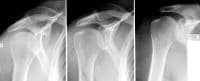 Normal plain radiograph of the shoulder in internal, external, and neutral positions.
 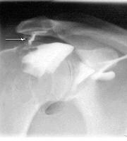 This
image depicts the channel between the articular capsule and the
subacromial-subdeltoid bursa in a complete rotator cuff tear.
This
image depicts the channel between the articular capsule and the
subacromial-subdeltoid bursa in a complete rotator cuff tear. 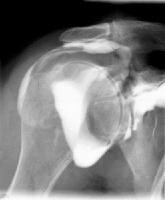 Even
if the channel cannot be always identified, the presence of contrast
medium in the subdeltoid-subacromial bursa signs the presence of a
complete rotator cuff tea
Even
if the channel cannot be always identified, the presence of contrast
medium in the subdeltoid-subacromial bursa signs the presence of a
complete rotator cuff tea
Normal plain radiograph of the shoulder in internal, external, and neutral positions. This
image depicts the channel between the articular capsule and the
subacromial-subdeltoid bursa in a complete rotator cuff tear. Even
if the channel cannot be always identified, the presence of contrast
medium in the subdeltoid-subacromial bursa signs the presence of a
complete rotator cuff tea
No comments :
Post a Comment